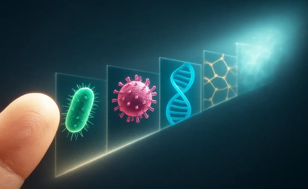
The intuitive idea of infinite “zoom” collides with physical limits: light diffracts, matter is damaged by high-energy probes, and spacetime itself may become undefined at extreme scales. This article builds from first principles to show where and why zooming in stops, and what it even means to look “after” atoms and nuclei.

The Optical Limit: Diffraction
Abbe’s Criterion and Airy Patterns
Optical microscopes are limited by diffraction: features closer than roughly half the wavelength of light blur together due to Airy disk overlap. Abbe’s criterion can be written as d≈λ2NAd≈2NAλ, setting a best-case lateral resolution near 200 nm with green light and high-NA oil objectives.
Practical Consequences for Biology
At ~200 nm, cells and many organelles are resolvable, while most viruses and macromolecular complexes remain below the limit. Label-free optical imaging struggles to reveal structures significantly under ~100–200 nm in conventional modalities.
Pushing Light Beyond Diffraction
Super-Resolution Modalities
Techniques such as STED, PALM, STORM, and structured illumination microscopy (SIM) circumvent the classical limit via nonlinear optics, stochastic localization, or patterned illumination and reconstruction. These methods can reach tens of nanometers under specific labeling, photophysics, and computational constraints.
Trade-offs and Use Cases
Super-resolution excels in cell biology when fluorescent labels and controlled conditions are available. However, it is not simple “zoom”: it demands specialized probes, careful dose management, and sophisticated data processing, and it remains orders of magnitude above atomic scales.
Switching Probes: Electrons for Atomic Scales
Why Electrons Resolve Atoms
Electrons have de Broglie wavelengths far shorter than visible light at typical accelerating voltages, enabling sub-nanometer—and even sub-ångström—resolutions in transmission electron microscopy (TEM/STEM). Aberration-corrected instruments routinely resolve atomic columns in crystals.
Three-Dimensional Reconstructions
Electron tomography and ptychography extend capability to 3D, with reconstructions approaching a few ångströms in suitable materials. Practical limits include radiation damage, sample thickness (tens of nanometers), vacuum compatibility, and stability.
Beyond Atoms: Quantum and Nuclear Scales
What an Atom “Looks Like”
Atoms are quantum objects: images at ångström scales reflect electron probability densities and scattering contrasts rather than hard surfaces. The nucleus itself is about femtometers in size (~10^-15 m), far beyond non-destructive microscopic imaging.
From Microscopy to Scattering
To probe subatomic structure, high-energy scattering replaces imaging. Particle accelerators infer internal structure from cross-sections and angular distributions, reconstructing models rather than producing direct pictures of intact matter.
The Conceptual Wall: Planck Scale
When Spacetime May Lose Meaning
The Planck length, about 1.6×10−351.6×10⁻³⁵ m, marks a scale where quantum gravity effects are expected to dominate. Heuristic arguments suggest that concentrating enough energy to probe below this would collapse the region into a micro black hole, making distances smaller than the Planck length operationally undefined.
Implications for “Infinite Zoom”
Approaching the Planck scale, frameworks like string theory or loop quantum gravity propose novel structure or discreteness. At this point, “zooming in” is no longer a meaningful visualization; instead, physics becomes speculative and fundamentally different in character.
Practical Summary by Scale
What Each Tool Can and Can’t Do
- Optical microscopy: best-case ~200 nm; live-cell friendly; label-free options limited at sub-100 nm.
- Super-resolution optics: tens of nanometers with labels and complex workflows; not atomic.
- Electron microscopy: to ~0.5 Å point resolution in materials; requires thin, stable specimens; destructive/dose-limited for soft matter.
- High-energy scattering: probes subatomic structure indirectly; no images of intact specimens.
- Planck scale: beyond current and foreseeable experimental reach; concept of distance may fail.
Comparison Table: Resolution, Requirements, and Limits
| Method | Typical Best Lateral Resolution | Sample Requirements | Strengths | Core Limits |
|---|---|---|---|---|
| Conventional optical (widefield/confocal) | ~200 nm | Minimal prep; compatible with live cells | Fast, accessible, non-destructive | Diffraction limit; poor below ~200 nm |
| Super-resolution (STED, PALM/STORM, SIM) | ~20–60 nm (mode-dependent) | Fluorescent labels; photostability; careful calibration | Sub-diffraction detail in cells | Complex, label-dependent, limited depth/speed |
| TEM/STEM (aberration-corrected) | ~0.5 Å point resolution | Ultrathin specimens; vacuum; beam-stable | Atomic columns, crystal lattices | Radiation damage; thickness; not for intact living systems |
| Electron tomography/ptychography | ~2–4 Å (context-dependent) | Tilt series; dose management | 3D near-atomic structure | Missing wedge, dose, reconstruction artifacts |
| High-energy scattering (HEP) | Sub-femtometer inference | Accelerators, targets/detectors | Probes subatomic constituents | No direct images; model-dependent |
| “Planck-scale” probes | Not achievable | — | Conceptual boundary | Physics may not allow operational meaning |
The Right Question at Each Scale
Matching Instrument to Phenomenon
“Zoom” is not universal; the relevant question is the characteristic length scale and allowable perturbation to the specimen. Choose optics for cellular dynamics, super-resolution for labeled nanoscale features, and electrons for atomic structure in robust materials.
What Comes After “After That”
Beyond atoms, microscopy yields to scattering and theory; beyond nuclei, only indirect inference remains; near the Planck length, even the notion of distance may break down. The end of zoom is thus both technical and conceptual.


